抗阻运动通过miR-21-5p/TSP-1途径改善老年男性增龄性循环内皮祖细胞功能损伤研究
作者: 韩书娜 孙一 叶琼
摘 要:观察规律抗阻训练对老年男性循环内皮祖细胞功能的影响并探讨miR-21-5p/血小板反应蛋白(TSP-1)在其间的可能作用机理。60名健康、无规律运动习惯的老年男性随机分为对照组和运动组,对照组维持日常生活习惯,运动组进行3次/周、共12周的抗阻训练干预。分别于试验前后,利用超声诊断系统测定肱动脉血流介导的血管舒张功能(FMD);取外周血分离培养内皮祖细胞,利用β-半乳糖苷酶染色法检测衰老水平,MTT比色法、Matrigel管腔形成实验分别检测体外增殖和成管能力,通过裸鼠颈动脉内膜拉脱模型检测在体内皮损伤修复能力,实时荧光定量PCR检测miR-21-5p和TSP-1 mRNA表达量,免疫印迹法测定TSP-1蛋白表达量;通过脂质体转染分别过表达miR-21-5p(miR-21-5p模拟物)和抑制TSP-1(TSP-1 siRNA)表达后,检测内皮祖细胞衰老和功能。结果:(1)训练依从性和安全性:运动组1例(3.3%)脱落,训练计划完成率为96.8%,无严重不良反应事件或心血管相关事件发生。(2)血管内皮功能和内皮祖细胞功能:试验后,与对照组比较,运动组FMD升高(P<0.05),内皮祖细胞β-半乳糖苷酶阳性细胞下降(P<0.05),体外增殖和成管能力增加(P<0.05),体内损伤血管再内皮化功能提高(P<0.05),miR-21-5p表达上调(P<0.05),TSP-1 mRNA和蛋白表达下调(P<0.05)。(3)细胞转染试验:转染miR-21-5p模拟物诱导miR-21-5p过表达后,内皮祖细胞TSP-1 mRNA和蛋白表达下降(P<0.05),体外增殖、成管能力以及体内损伤血管再内皮化功能改善(P<0.05),然而,β-半乳糖苷酶阳性细胞无显著性变化(P>0.05);转染TSP-1 siRNA诱导TSP-1沉默后,β-半乳糖苷酶阳性细胞下降(P<0.05),体外增殖、成管能力以及体内损伤血管再内皮化功能提升(P<0.05)。结论:抗阻训练通过上调miR-21-5p抑制TSP-1表达来改善老年男性内皮祖细胞功能,通过下调TSP-1延缓内皮祖细胞衰老,因此miR-21-5p和TSP-1是抗阻训练发挥心血管保护效应的重要靶点。
关 键 词:运动生物化学;抗阻训练;内皮祖细胞;血管内皮功能;老年男性
中图分类号:G804.7 文献标志码:A 文章编号:1006-7116(2025)01-0150-07
A study on resistance training improves age-related circulating endothelial progenitor cell functional impairment in old males by miR-21- 5p/TSP-1 pathway
HAN Shuna1,SUN Yi2,YE Qiong3
(1.Department of Physical Education,Sichuan International Studies University,Chongqing 400033,China;
2.School of Physical Education,Jilin University,Changchun 130012,China;
3.Department of Physical Education,Chongqing Preschool Education College,Chongqing 404000,China)
Abstract: To observe the effect of regular resistance training on the function of circulating endothelial progenitor cells in elderly men and to explore the possible mechanism of miR-21-5p/thrombospondin 1 (TSP-1). 60 healthy old males with no regular exercise habits were randomly divided into control group and exercise group. The control group maintained their daily habits, and the exercise group underwent resistance training intervention three times per week for a total of 12 weeks. Before and after the experiment, an ultrasonic diagnostic system was used to measure brachial artery flow mediated dilation (FMD). Peripheral blood was taken to isolate and culture endothelial progenitor cells, β-galactosidase staining to detect the aging level, MTT colorimetry and Matrigel tube formation assay to detect the proliferation and tube-forming ability in vitro respectively, the repair ability of endothelial damage in vivo through the nude mouse carotid artery intimal pull-off model, real-time fluorescence quantitative PCR to detect the expression of miR-21-5p and TSP-1 mRNA, and the expression of TSP-1 protein by Western blotting, were determined. After overexpressing miR-21-5p (miR-21-5p mimic) and inhibiting TSP-1 (TSP-1 siRNA) expression respectively by lipofectamine transfection, endothelial progenitor cell senescence and function were detected. The results show that: (1) Training compliance and safety: 1 case (3.3%) dropped out, the training plan completion rate was 96.8%, and no serious adverse events or cardiovascular-related events occurred in the exercise group. (2) Vascular endothelial function and endothelial progenitor cell function: After the experiment, compared with the control group, FMD increased (P<0.05), β-galactosidase-positive cells of endothelial progenitor cells decreased (P<0.05), the proliferation and tube formation abilities in vitro were increased (P<0.05), the reendothelialization function of injured blood in vivo was improved (P<0.05), the expression of miR-21-5p was up-regulated (P<0.05), and the expression of TSP-1 mRNA and protein was down-regulated (P<0.05) in the exercise group. (3) Cell transfection test: After transfection with miR-21-5p mimic to induce the overexpression of miR-21-5p, the expression of TSP-1 mRNA and protein in endothelial progenitor cells decreased (P<0.05), the proliferation and tube formation ability in vitro and reendothelialization function of injured blood in vivo were improved (P<0.05), however, there were no significant changes in β-galactosidase-positive cells (P>0.05). After transfection of TSP-1 siRNA to induce TSP-1 silencing, β-galactosidase-positive cells decreased (P<0.05), the proliferation and tube formation ability in vitro and reendothelialization function of injured blood in vivo increased (P<0.05). The conclusion reveal that: resistance training inhibits TSP-1 expression by up-regulating miR-21-5p, improves endothelial progenitor cell function, and delays endothelial progenitor cell aging by down-regulating TSP-1 in elderly men. Therefore, miR-21-5p and TSP-1 are important targets for the cardiovascular protective effects of resistance training
Keywords: sports biochemistry;resistance training;endothelial progenitor cells;vascular endothelial function;old males
增龄可导致血管内皮功能下降,从而诱导血管内膜层增生,最终引发多种心血管疾病[1]。血管内皮细胞通过释放血管活性物质调节血管的舒缩活动,然而各种原因导致内皮细胞受损后,易发生动脉粥样硬化、血管狭窄、堵塞等病理性改变[2]。由于损伤后内皮细胞增殖和修复能力有限,骨髓中的内皮祖细胞动员入血并诱导分化为内皮细胞以修复受损血管[3]。Dight等[4]发现衰老导致循环内皮祖细胞基线数量减少以及功能降低,内皮祖细胞修复和更新受损血管的能力下降。
运动疗法是防治心血管疾病的重要策略[5-6]。有氧运动对增龄所致心血管损伤的有益效应以及内皮祖细胞的作用机制已得到广泛证实[7-13]。抗阻训练作为老年人运动处方的重要方式,具有增加肌肉含量、预防肌萎缩,提高骨密度和肌肉力量,预防跌倒与骨折[14],同时通过释放多种肌肉因子(如鸢尾素、白细胞介素-6等)调节远隔器官(包括心血管系统、骨骼系统)的功能[15-16],因此同样具有心血管保护作用,然而其是否通过内皮祖细胞发挥健康效应仍未确定。据报道,衰老过程中肌肉含量下降(肌少症)导致肌肉因子(如鸢尾素、白细胞介素-6等)释放减少,从而间接影响内皮祖细胞功能,而外源性补充鸢尾素可增加骨髓内皮祖细胞数量[17]。相较于有氧运动,抗阻训练能够促进肌肉因子大量分泌[15],推测其理应对内皮祖细胞数量和功能产生有益作用。
微小核糖核酸(miRNA)是一类由内源基因编码的长度约20~24 bp的非编码单链RNA,通过调控转录后的基因表达参与细胞分化、发育、组织生长等诸多生理过程[18]。miR-21-5p广泛存在于哺乳动物组织和器官中,对心血管功能起重要的调节作用。Mayourian等[19]发现,人骨髓间充质干细胞分泌的外泌体富含miR-21-5p,能有效增强心肌收缩力。Souza等[20]研究证实,运动上调心力衰竭小鼠内皮祖细胞miR-21-5p表达量,提示运动可能通过调节miR-21-5p表达改善内皮祖细胞功能。血小板反应蛋白(Thrombospondin 1,TSP-1)是内源性血管生成抑制因子,对细胞衰老具有促进作用[21]。研究发现,增龄可诱导组织(尤其是心肌)中TSP-1表达上调,进而引起细胞功能减退并加速衰老进程[22]。进一步研究证实,TSP-1表达上调可能导致内皮祖细胞功能和血管生成能力下降[23]。然而,衰老促进TPS-1表达与内皮祖细胞功能降低之间的关系尚未厘清。Hu等[24]发现,内皮祖细胞经由外泌体途径将miR-21-5p转运至内皮细胞,miR-21-5p通过下调TSP-1表达改善内皮细胞功能。据此推测,miR-21-5p/TSP-1信号通路可能参与运动对内皮祖细胞功能的调节。因此,本研究旨在观察规律抗阻训练对老年男性循环内皮祖细胞功能的影响并探讨miR-21-5p/TSP-1在其间的可能作用机理,为运动抗衰老效应提供理论基础、有效方法和干预靶点。
1 研究对象和方法
1.1 研究对象
1)样本量估算。
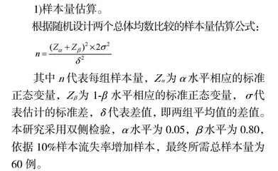
根据随机设计两个总体均数比较的样本量估算公式:
其中n代表每组样本量,Zα为α水平相应的标准正态变量,Zβ为1-β水平相应的标准正态变量,σ代表估计的标准差,δ代表差值,即两组平均值的差值。本研究采用双侧检验,α水平为0.05,β水平为0.80,依据10%样本流失率增加样本,最终所需总样本量为60例。
2)研究对象
招募重庆市某社区60~75岁老年男性60名。纳入标准:(1)身心健康;(2)无规律运动习惯;(3)自愿参加本次研究,并且能坚持配合运动者。排除标准:(1)患有心脑血管、代谢和肾脏等疾病;(2)运动风险筛查为中、高危者;(3)骨骼肌肉系统急性慢性损伤及运动障碍者;(4)认知障碍;(5)长期服药史以及吸烟酗酒等不良习惯者。所有人员均签署知情同意书,本研究获得吉林大学伦理委员会批准(NO.2023030701)。按照随机数字表法将受试者分为对照组和运动组,两组受试者入组前在年龄、身高、体重、体质量指数、安静心率、血压、心血管疾病家族史等参数间比较,差异均无统计学意义(P>0.05),组间具有可比性。
1.2 实验方法
对照组维持日常生活习惯,运动组进行12周渐进性抗阻训练。分别于试验前后,利用超声诊断系统测定肱动脉血流介导的血管舒张功能(flow mediated dilation,FMD);取外周血分离培养内皮祖细胞,利用β-半乳糖苷酶染色法检测衰老水平,MTT比色法、Matrigel管腔形成实验分别检测体外增殖和成管能力,通过裸鼠颈动脉内膜拉脱模型检测在体内皮损伤修复能力,实时荧光定量PCR检测miR-21-5p和TSP-1 mRNA表达量,免疫印迹法测定TSP-1蛋白表达量;通过脂质体转染分别过表达miR-21-5p(miR-21-5p模拟物)和抑制TSP-1(TSP-1 siRNA)表达后,检测内皮祖细胞衰老和功能。
(1)血管内皮功能测定。利用彩色超声诊断系统(LOGIQ7型,美国GE公司)测定肱动脉FMD反映血管内皮功能。受试者取仰卧位,以右侧肘横纹上5 cm肱动脉段为靶血管。先测量静息时的肱动脉舒张末期内径D0,随后通过血压计袖带加压至200 mmHg完全阻断血流5 min后迅速放气,测定肱动脉舒张末期内径D1。用反应性充血前后肱动脉内径的变化率计算FMD(%)=(D1-D0)÷D0×100%。
(2)外周血内皮祖细胞培养和鉴定。内皮祖细胞培养:分别于试验前后清晨空腹状态下肘正中静脉取血20 mL,肝素抗凝。Ficoll密度梯度离心法获取外周血单个核细胞。用M199培养基置于37℃、5%CO2的细胞培养箱中孵育,利用倒置显微镜(DM IL LED,德国徕卡公司)观察细胞形态。内皮祖细胞鉴定:利用吞噬实验鉴定内皮祖细胞。将单细胞悬液与Dil标记的乙酰化低密度脂蛋白(acetylated low density lipoprotein,ac-LDL)(Dil-ac-LDL)(20 μg/mL)溶液于37℃孵育120 min,4%多聚甲醛固定15 min,PBS洗涤后与FITC标记的荆豆凝集素1(ulex europaeus agglutinin-1,UEA-1)(FITC-UEA-1)(10 μg/mL)溶液中于37℃孵育60 min,甘油密封。激光共聚焦显微镜(TCS SP8 X,德国徕卡公司)下随机选取5个视野,计数双染细胞数(即内皮祖细胞)占总细胞数的百分比。